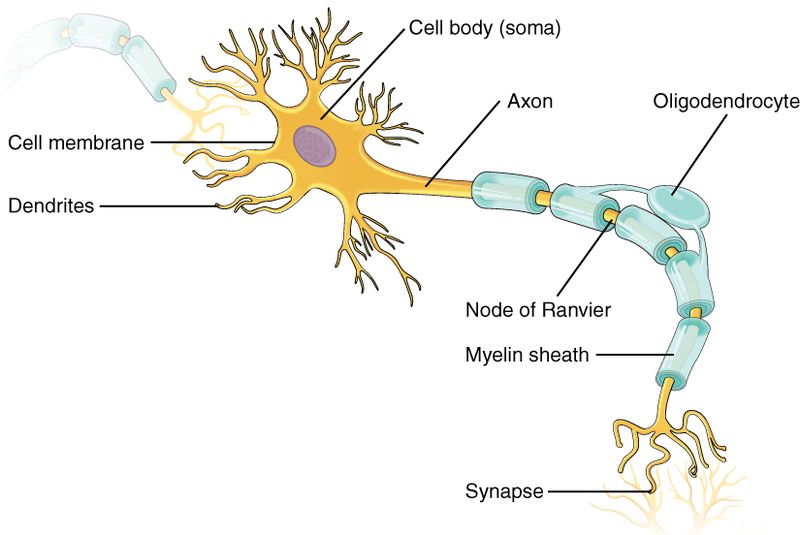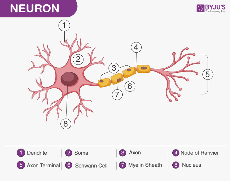

(LeDoux, 42) A critical part of the neuron.

The result is that messages sent out from one cell can affect many others. However, each axon branches many times before it ends, allowing a single neuron to spawn many terminals. (Kandel, 64) Most neurons have only axon. Often splits into one or more branches along its length. (The Brain-Francis Crick, 132) Emerges at one end of the cell body and can extend up to several feet. (Goldberg, 39) Long fiber of the neuron, often with thousands of contact points. (LeDoux, 40) A protrusion emanating from the cell body that along with another neuron’s “dendrite” makes possible the connections between neurons. (Ramachandran, 9) Axons are "output" channels and dendrites are "input" channels. The axon’s function is to send data out of the cell. A projection from the neuron "cell body” that can reach long distances in the brain. This study continues our laboratory's work in discerning the function of DPP6 and here provides compelling evidence of a direct role of DPP6 in Alzheimer's disease.Axon: one of the three main (components) of the neuron. Together these results indicate that aging DPP6-KO mice show symptoms of enhanced neurodegeneration reminiscent of dementia associated with a novel structure resulting from synapse loss and neuronal death. We finally show that aging DPP6-KO mice display circadian dysfunction, a common symptom of Alzheimer's disease. And levels of proinflammatory or anti-inflammatory cytokines increased in aging DPP6-KO mice.

The amyloid β and phosphorylated tau pathologies were associated with neuroinflammation characterized by increases in microglia and astrocytes. Aging DPP6-KO hippocampi contained fewer total neurons and greater neuron death and had diagnostic biomarkers of Alzheimer's disease present including accumulation of amyloid β and APP and increase in expression of hyper-phosphorylated tau. We used in vivo MRI to show reduced size of the DPP6-KO brain and hippocampus. Here we also found evidence that aging DPP6-KO mutants show additional changes related to Alzheimer's disease. In this current study, using immunofluorescence and electron microscopy, we confirm that both APP and amyloid β are prevalent in these structures and we show with immunofluorescence the presence of similar structures in humans with Alzheimer's disease. These novel structures appear as clusters of large puncta that colocalize NeuN, synaptophysin, and chromogranin A, and also partially label for MAP2, amyloid β, APP, α-synuclein, and phosphorylated tau, with synapsin-1 and VGluT1 labeling on their periphery. Recently, we have described novel structures in hippocampal area CA1 in aging mice, apparently derived from degenerating presynaptic terminals, that are significantly more prevalent in DPP6-KO mice compared to WT mice of the same age and that these structures were observed earlier in development in DPP6-KO mice. DPP6-KO mice are impaired in hippocampal-dependent learning and memory and exhibit smaller brain size. We have previously reported that the single transmembrane protein Dipeptidyl Peptidase Like 6 (DPP6) impacts neuronal and synaptic development. Recent studies with single-molecule imaging in live neurons confirmed and extended the role of Arc mRNA trafficking in synaptic plasticity. Therefore, Arc mRNA inter- and intramolecular trafficking is essential for the modulation of synaptic activity required for memory consolidation and cognitive functions. Such complex can be loaded into extracellular vesicles and transported to other neurons or muscle cells carrying not only genetic information but also regulatory signals within neuronal networks. Arc can form a capsid-like structure that encapsulates a retrovirus-like sentence in the 3′-UTR (untranslated region) of Arc mRNA. Therefore, Arc might evolve through “domestication” of retroviruses. The structure of Arc is similar to the viral GAG (group-specific antigen) protein, and phylogenic analysis suggests that Arc may originate from the family of T圓/Gypsy retrotransposons. Arc mRNA is exported into the cytoplasm and can be trafficked into the dendrite of an activated synapse where it is docked and translated.

Transcription of the Arc gene may be induced by a short behavioral event, resulting in synaptic activation. Activity-regulated cytoskeleton-associated protein (Arc) is a master regulator of synaptic plasticity and plays an important role in controlling large signaling networks implicated in learning, memory consolidation, and behavior. Intracellular localization of a protein can be achieved by mRNA trafficking and localized translation. Synaptic activity mediates information storage and memory consolidation in the brain and requires a fast de novo synthesis of mRNAs in the nucleus and proteins in synapses.


 0 kommentar(er)
